Advanced Family Eye Care administers a wide array of tests and procedures to examine your eye health and visual acuity. These tests range from basic ones; like reading from a classic eye chart, to complex tests where we use high-powered lenses to visualize the finite anatomical structures of your eye. At AFEC we pride ourselves at always being on the frontline of new technological developments and bringing those technologies to our patients.
Depending on the complexity of the test required, a comprehensive exam can take up to an hour to fully evaluate your vision and the health of your eyes.
Here are eye and vision tests that you are likely to encounter during a routine comprehensive eye exam:
The first tests typically administered during a comprehensive eye exam are visual acuity tests. This involves reading from a projected eye chart that is designed to measure the sharpness of your vision from afar, while a smaller chart tests your visual prowess from up close.
Color blindness is something eye doctors will try to rule out in the early stages of a visual acuity test. This includes the common form of the condition, which is often hereditary, as well as color blindness brought about by unrelated health problems.
Having strong visual acuity is important, but only if both eyes can complement one another, which is why the cover test is designed to determine how well your eyes work as a pair when focusing on an object.
This type of test can identify subtle binocular discrepancies or “Strabismus”, which can lead to eye strain or “Amblyopia”, otherwise known as lazy eye. This involves focusing on objects, one eye at a time, from both near and far.
Our providers will then determine whether the uncovered eye must move in order to fixate on the target. If it does, this could indicate strabismus or a more subtle binocular vision problem that may lead to eye strain or amblyopia.
Our providers will likely perform this test early on to obtain an approximation of your eyeglass prescription.
For this test, the lights in the exam room are dimmed. You’re then asked to fixate on a large target in the room (often the large “E” at the top of the exam chart). As you do this, our providers will direct a beam of light toward your eye while placing different lenses in front of them. Depending on how the light reflects from your eye, our providers can typically develop an approximate guess of what your prescription should be. The results are surprisingly accurate at times and can be especially helpful when testing the vision of infants since no dialog is necessary.
When determining your preferred prescription, our providers will conduct a test that is known as a refraction test. For this exercise, an instrument known as a “phoropter” is placed in front of your eyes, while a series of different lenses are cycled through. The purpose of this is to identify which lens provides you with the best focus. As you do this, our providers will ask you a series of questions to help fine-tune the lens selection. This enables our providers to identify the “perfect” prescription for you based on your response to the test.
Refraction allows our providers to determine your level of hyperopia (farsightedness), myopia (nearsightedness), astigmatism, and presbyopia.
Our providers routinely use an autorefractor to automatically determine a prescription. It can save time and replace Retinoscopy entirely.
For this test, your head is stabilized by a chin cup while you focus on a pinpoint of light or some type of image. This is designed to help identify the lens power required to accurately focus light on your retina. Autorefractors are especially useful for young children who may not sit still, pay attention or interact with the eye doctor during a Retinoscopy.
Published reports have indicated that modern Autorefractors are not only very accurate but save a great deal of time since autorefraction takes only a few seconds. The results obtained from the automated test greatly reduce the time required to perform a manual refraction and determine your eyeglass prescription.
A contact lens has the ability to alter or damage the physiological structure of the cornea (the front of your eye). As a result, a topography is strongly recommended for anyone who plans to wear contact lenses.
Much like a topographical survey of the earth, a corneal topography gives our providers a map of the microscopic hills and valleys on the front surface of the eye (cornea).
At AFEC we use a very advanced Topographical mapping system called a Humphrey Atlas. This is a very simple test to perform. The patient places their chin on a chin rest and looks into the instrument one eye at a time staring into a fixation target to stabilize the eye. Then a light is reflected off of the surface of the eye while the instrument calculates the map.
This data can then be used to monitor unwanted tissue changes as a result of contact lens wear or other inherited corneal pathology such as Keratoconus. This data is also invaluable for selecting the proper contact lens for a patient.
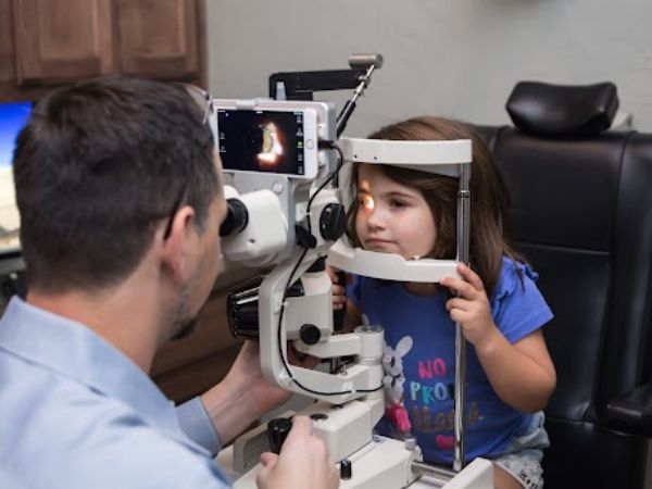
In order to determine overall eye health, an instrument known as a slit lamp is used to obtain a highly magnified view of the structures in each eye. This affords our providers the opportunity to thoroughly evaluate your eye health and detect any signs of infection or disease.
Much like an Autorefractor, your head is stabilized by resting it atop a chin cup. A light is then focused on your eye. Our doctors look through a set of oculars (much like a microscope in a science lab) and examine each part of your eye in turn.
They will first examine the structures of the front of your eye (lids, cornea, conjunctiva, iris, etc.). Then, with the help of a special high-powered lens, they will view the inside of your eye (retina, optic nerve, macula, and more).
A wide range of eye conditions and diseases can be detected with slit-lamp examination, including cataracts, macular degeneration, corneal ulcers, diabetic retinopathy, etc.
There are a variety of ways to administer a Glaucoma test, which measures the pressure inside your eyes in order to achieve a diagnosis. A Tonometer is used to diagnose the condition. One example is the “puff of air” test, technically known as the no-contact tonometer or NCT. You first stabilize your head by resting it atop a chin cup. Our providers or a qualified assistant will then have you look at a light inside the machine while they direct a puff of air at the corneal surface of your eye. It’s completely painless and based on your resistance to the puff of air, they can determine your intraocular pressure (IOP). If you have high eye pressure, you may be at risk for, or have glaucoma.
Due to the unpopularity of the “puff test”, AFEC uses the latest tonometer known as the ICARE Tonometer. This test is much more patient-friendly and more accurate than the “air puff”
Another type of glaucoma test involves an instrument known as an applanation tonometer. This is the gold standard of tonometer testing. For this, our doctors will put yellow eye drops in your eye to numb it. Your eyes will feel slightly heavy when the drops start working. This is not a dilating drop — it is a numbing agent combined with a yellow dye that glows under blue light. They will then ask you to stare into the slit lamp while they gently touch the surface of your eye with the tonometer to measure your IOP. Like the NCT, an applanation tonometer is painless. You may feel the tonometer probe tickle your eyelashes, but the whole test is completed in just a few seconds.
Patients typically have little or no warning of glaucoma until they have experienced significant vision loss. For this reason, routine eye exams that include a tonometer test are essential to rule out early signs of glaucoma and protect you from vision loss.
For certain kinds of tests, our providers will need to examine the internal structures of your eye. In order to accomplish this, they may administer a dilating drop to enlarge your pupils. Dilating drops usually need 20 to 30 minutes before taking full effect. During dilation, you will be especially sensitive to light (since more light is entering your eye) and you may notice difficulty focusing on objects up close. These effects can last for several hours, depending on the strength of the drop used. Pupil dilation is very important because it affords our doctors the most thorough evaluation of your internal eye health.
Once the drops have taken effect, our providers will use various instruments to look inside your eyes. You should bring sunglasses with you to your eye exam, to minimize glare and light sensitivity on the way home. If you forget to bring sunglasses, the AFEC staff will give you a disposable pair for your convenience.
AFEC routinely utilizes retinal imaging. Because of this in most instances, sufficient information can be obtained about the retina which reduces the need for dilation.
In some cases, our doctors may want to check for the presence of blind spots (scotomas) in your peripheral or “side” vision. They do so by performing a visual field test. Blind spots can originate from eye diseases such as glaucoma. Performing an analysis of your blind spots may also help identify specific areas of brain damage caused by a stroke or tumor. At AFEC we utilize a very advanced Automated Visual Field device called a Humphrey Matrix. This instrument is among the fastest and most accurate at testing “side” vision.
The above list represents the most common testing performed at AFEC. However, if our doctors detect an abnormality, there are many more tests that they can utilize to ensure you receive a thorough exam.


